Friday, December 2, 2011

A great experience

My vision therapist set up something really cool for me. She has another adult strabismic in therapy and she set up a time for us to meet and do a 2 hour block of vision therapy together. This other patient, let's call her... Tara, has had 8 surgeries, has been both an esotrope and an exotrope (currently eso), is a pretty severe amblyope in one eye, and  has double vision 100 percent of the time. My vision therapist thought it would be good if we got together, because she said we both "speak the same language" and might be able to help each other.

Finally, this Friday, we got a chance to meet and play around with some red/green stuff! It was great. It turns out that Tara's double vision allows her to "cheat" on most red/green exercises, and so far it has been very difficult for her to get any fusion or overlapping of the two doubled images that she sees. The images just don't want to overlap and make a combined picture, and she doesn't know how to move her eyes to move the doubled images over top of each other.

I started to get so interested in what was going on with Tara's double vision that I spent almost the whole time asking her questions and trying different things with her. One of the toys at the VT office was this ancient looking device that looks like two flashlights connected to box, with one flashlight projecting a red circle and one flashlight projecting a green dot.

When my vision therapist pointed the flashlights at the wall and put the green dot inside the red circle, Tara could not see the green dot inside the red circle. She saw the green dot to the left and under the red circle. When we were doing other red/green activities, she also mentioned that she saw double images that were vertically displaced.

 
From what I've read and heard, I seem to remember that vertical misalignment is harder to treat than an exo or eso deviations. If one eye is wandering out horizontally, then it is possible to learn how to pull it in. But if one eye is wandering up, it can be difficult to learn how to pull it down. Or so I've heard. I've also heard that if the vertical deviation isn't too extreme, the brain can compensate and fuse the images, but if it's too far, then it can be really hard.

I thought that maybe if she used a vertical prism, she would have more luck with fusion. I'm not sure if it's frowned upon to use prisms to help with vertical deviations, or if you're just supposed to "deal with it" until you suddenly have the eye strength to do it yourself, but I wanted to try it. So I ran and grabbed the box of loose prisms and found that a 6 prism diopter base down over one eye (or base up over the other eye) allowed her to see the dot in the circle. One image, no vertical displacement. Cool!!!!


We also did some other stuff and I tried out some ideas for physical diplopia practice. And then the three of us started brainstorming and came up with a neat idea for an exercise we want to develop involving black, red, and green felt and Nerf guns (details to come later). It was really fun and inspiring experience all around! I'm more inspired to do my own homework now, and it even made me consider maybe going to optometry school some day. Vision therapy is just so fun!

Sunday, October 23, 2011

I'm back!

After 7 months, I have returned. My personal life became kind of crazy for a while (a serious car accident, among other things) and I kind of forgot about vision therapy. At that time, seeing out of one eye was good enough for me. But everything is okay now! I'm back in vision therapy, and I have even more ideas and inventions floating around in my head than before.

If you tried to email me, I'm sorry that I didn't answer. I will slowly try to go through my old emails and see what I missed. Thanks :)

To prove that I am really back, here is a picture of me doing one of my first back-in-vision-therapy homework assignments: wear the red/green glasses and track the red/green ball as it floats around your face. Thankfully, I'm finding that I can still flat fuse red/green stuff like that. It seems that I haven't lost any of the talents that I gained from the first round of vision therapy, but I sure do feel a little rusty.

Wednesday, March 30, 2011

Just a little

I saw a little depth at my last vision therapy appointment. For the first time! Just a little! But still... it happened! Here's the story...

In Effective Strabismus Therapy, Dr. Israel Greenwald talks a lot about working with strabismus patients to achieve red/green luster while looking at a white wall. The patient wears red/green glasses, stands up close and stares at a big empty while wall, and tries to make the colors intermix and become inseparable. To help achieve this, he recommends that the patient wear high plus lenses (+15 to +20). These high plus lenses are basically superpowered magnifying glasses that make everything blurry. This removes all figure/ground distinctions and allows for easier fusion.

I asked my vision therapist if we could get some high plus lenses and she said 'Sure!' She called in an order to the lab, and I thought everything would be fine. It turns out, though, that it's very hard to find a place that even makes plus lenses that strong. The strongest that I have seen available for order on the internet are +12. (+12 is actually very strong though and would probably work, but that's not part of the story.)

So last Friday, I brought up the topic of plus lenses again and my vision therapist had an idea. There were lots of contacts in the office, so why not try high plus contacts? That way, there wouldn't even be a chance to "peek around" the glasses and ruin the blurring between figure and ground. They weren't any +20, but there were some +6 available. (or maybe they were +4, I can't remember).

So I said yes, even though I was kind of terrified. (You remember what happened last time someone got all up in my eyes.) And it turns out, contacts are really hard to get in! I did the usual - hold your eyelids down like they are trying to escape - type of thing, but then I would involuntarily try to shut my eye so hard that my eyelids would turn inside out and block the contact anyway. It was both funny and gross.

20 minutes later, when they were finally in, I got to experience the world as myope (near sighted person). I actually found it quite nice. My eyes couldn't focus on anything and thus didn't try - it felt rather relaxing. It also blurred out everything past my nose, leaving me encapsulated in a blurry womb. I've always been an anxious and shy person, wondering what people are thinking about me (are they looking at my crooked eye??), and for once, I couldn't see anybody's faces. I just had to assume that everyone was smiling at me. I wouldn't want to live with uncorrected blurry vision, of course, but it was nice to take a vacation in blurry land for a while.

So we tried some red/green luster with the white wall, and the extra plus seemed to help (more plus would have been better, though). While, the contacts were in, we also tried some other things. I had some vague memories of reading that dim light can help fusion and help break suppression, and something about contrast levels.... and something about ARC and peripheral fusion... and something about something else... So we decided to try out some polarized vectograms while I was wearing the plus contacts, just for fun, and.... something happened!

First, I was looking at this "figure 8" vectogram:


I was looking at this one the previous week, and while rocking side to side, I noticed that one of the little triangles was moving with me. It only did it for a second, and I couldn't really get it to happen again, but I knew that this vectogram was doing something funny to my brain. So, while wearing the blurry contacts, I decided to start with this one.

I put the polarized glasses on and started to rock side to side like last time. And suddenly little triangles started popping off the screen! They didn't stay popped out for very long, but it happened more than a few times, especially (for some reason) on the triangles on the left hand side of the picture.

It reminded me of those pop-up story books - the kind where you pull a tab and little paper figures extend off the page. It was as if someone was pulling a tab behind the vectogram and making the triangles pop out. At one point, I even saw a wave of depth undulate across the vectogram and then disappear. It was pretty neat.

Excited, I tried another vectogram - the one my vision therapist calls "creepy clown."


Again, something happened! Nothing really popped off of the screen, like those little triangles did in the figure 8 vectogram.. It was more like... the screen fell away and I was looking through a portal into another dimension... A dimension of scary living devil clowns!

The area with the most depth was that area between creepy clown's legs, where you can see his two legs, his hand, and the stool. It's was very depthy in there. And yes, I do appreciate how creepy it is to have one of my first experiences with depth involve looking between a clown's legs. Some optometrist out there sure has a sick sense of humor.

Also, every time the screen fell away and got depthy, I got the strange sensation that someone was punching me in the stomach. That was pretty neat, too.

So that's the story. It wasn't in "real life", and it was blurry and unstable. But it HAPPENED. Up until this point, I have been secretly fearing that maybe I was born without those special binocular neurons. Maybe I became strabismic because my brain was missing some pieces... Now I know that's not the case. I just need to keep practicing.

Monday, March 21, 2011

What is "fusion"?

I'm writing this entry to describe my experience with "fusion" in vision therapy. I'm putting "fusion" in quotes, because I'm still not quite sure if what I am experiencing is fusion, or even what the definition of fusion really is. I know that, basically, fusion means two eyes making one picture. But what I've seen and noticed seems much more complex, and involves brain processes that I can't even begin to understand. In any case, this is what I have noticed about fusion so far...

First, I have noticed that fusion is not just one process. There seem to be several "types" of fusion that I have come across, and even within these "types", lines get blurred and things get murky. These types may even exists on a continuum. However, I will try my best to explain what I have experienced. Here are the three types of fusion that I have come up with:

1. Cut and paste
 This is the easiest kind of fusion for me. The images from each eye are selectively cut and pasted together into one image through some brain photoshoppery. An example of this would be the anti-suppression playing cards that I love so much.


When viewed through red/green(blue) glasses, these are the images that the brain must combine:


How does the brain do this? In my subjective experience, it feels as if my visual system is using scissors and glue to make a sort of collage.


Of course, it doesn't look like that. But it feels as if that's the process that my brain is going through - choosing the important elements from each eye, and deciding which to use in the final "collage." If the brain did not go through such a process of defining importance and cherry picking input, then what would stop the brain from making an image such as this?


The brain would not make the above picture, and one would never see the cards all blank like that. I dare you to try! The brain looks for meaningful input, and assembles everything together accordingly. That is why, when making anti-suppression activities, I am always writing letters on everything. I have learned from the playing cards that the brain cannot ignore letters.

For me, this type of fusion becomes more difficult as finer and more precise "brain cuts" and "brain pastes" are required. For example, fuse - by convergence or divergence - the image below:


What you should see is something like this:


This is quite hard for me to do, and I believe I know the reason why. Look at all of the places where the lines are in conflict (in pink). This would require some very creative cutting and pasting.


In fact, this comes close to requiring the brain to create a completely new image (rather than a collage), which brings me to the next kind of fusion.

2. Transmutation
In this type of fusion, something new is created that neither eye can see alone. This is not a composite of two images, as is the case with cut and paste fusion. Two images are combined in such a way that they create a new, unique third image.This is certainly not a collage. It's a new painting!

An example would be the "luster" effect. By convergence or divergence, fuse the two images below:


What you should see is one grid in the middle, with the red square and the yellow square magically transmuted into a new color. This new color was not perceived by either eye, it was created in the brain. (I imagine it will be glowing like it's fresh out of a nuclear reactor.)


This type of fusion is exceedingly difficult for me, and I'm not sure that I have properly achieved it. When I try to do luster activities, I tend to get flashing, alternating between colors, rather than the creation of a new color. I also imagine that stereoscopic vision requires a great deal of transmutational fusion, and thus it is very important that I get better at it! I can't be satisfied with merely cutting and pasting; my brain needs to learn how to blend and create.

3. Stereopsis
What happens here? I'll let you know when I see it. I've heard that salads, tomatoes, horse skeletons, steering wheels, and coat sleeves all start jumping out to say hello. It sounds pretty awesome.

Thursday, March 17, 2011

My left eye - a VT update

(I've lost track of the weeks. They're just flying by!)

The theme for my vision therapy lately has been "back to basics." My vision therapist and I were talking, and we decided that we skipped over some important stuff in the beginning. With new patients, she normally starts out with a lot of monocular patched activities, tracking, pursuits, body bilaterality, and so on. But in my case, I was very eager and kept being like, "Wow! Is that a cheiroscope? I read about that. Can I take that home?" And so we got a little ahead of ourselves.

As I've been progressing and starting to really get a feel for my eyes, I've been realizing the importance of those beginning foundational activities. I'm becoming very aware of how unequal my eyes are, and I can see how much of a hindrance that can be. Therefore, for the past few weeks, we've been going back to those basic beginning activities.

Specifically, I've been working on my left eye - the amblyopic and frequently turned eye. Here is what I have noticed about it:

1. It is terrible at pursuits, especially going from right to left. It prefers a quick succession of saccades, or what Dr. Len Press told us are "catch-up ballistic saccades." My eye thinks that it's tricking me with these saccades, and that I won't notice that they aren't real pursuits. You're wrong, eye!

2. It has trouble knowing where it's pointing. I sometimes think that I am using the center of my eye to track something, but really I have stopped moving my eye (because the object has moved to a place where it's hard to move my eye smoothly) and I'm just seeing it my peripheral vision. My eye feels like that is "good enough" and doesn't realize that it's not even tracking anymore.

3. It doesn't like to initiate movement or be the leader. It just wants to follow what my right eye is doing.

4. It has trouble maintaining a stable gaze. When I try to fixate on something for more than just a second, my eye jitters around the target. I've witnessed this using the MIT and seeing the Haidinger brush dance around the target that I was (trying to) fixate on.

5. It's even nystagmus-y on occasion. Sometimes when I wake up in the morning and my right eye is occluded by the pillow, I will look at the alarm clock with my left eye and the image of the clock will jerk back and forth over and over.

(and I'm sure I'll discover more as time goes on)

Amazingly, I had no awareness of any of this until very recently. How could I not notice all these years?

Now that I have noticed, I have been busy working away on my monocular skills with activities like:

tracking / pursuits with the monster
word searches with progressively smaller letters
marsden ball tracking
MIT mazes
tracking while keeping something in a circular afterimage
and so on...

And of course I've been throwing in some physiological diplopia and antisuppression exercises, because life would be boring with without them.

Monday, March 14, 2011

Thoughts on esotropia (and scotomas)

Sometimes, I lay awake at night, thinking of esotropia and how mysterious it is. I've read and heard over and over that exotropia is easier to treat than esotropia. Good thing I'm exotropic, I think to myself. But then I remember that I used to be very esotropic. I was a cross eyed baby! I had congenital/infantile esotropia. That's how this whole strabismus mess started.

I developed exotropia after my first surgery, and then a second and third surgery couldn't get rid of it. Is it really true that, because my strabismus surgeries accidentally made me exotropic, I now have a better prognosis than my esotrope friends? Even though my original condition was esotropia? That seems weird to me. Or lucky. Or something.

Perhaps it's all a mystery, and nobody knows...

After my previous post on suppression scotomas, I got interested in drawing out the suppression scotomas of esotropes. So here goes.


Both eyes are able to see the diamond, one centrally and one peripherally, and double vision should result because the eyes are not aligned. However, only one diamond is seen, because the peripheral diamond image is falling on a suppression scotoma.

In my previous post, I also mentioned that I had a habit of peeking around my suppression scotoma by overconverging. Is it possible for an esotrope to peek around their suppression scotoma? Yes!

If both eyes overdiverge and fixate on a point behind the diamond, that would make the diamond image fall outside of the suppression scotoma (and the image would double). However, this is easier said that done. I have trouble diverging my eyes at will, and I can imagine that an esotrope would have the same difficulty. After all, if they could diverge their eyes like that, they wouldn't be cross eyed! So it's possible that this may not happen as often.

It does explain something interesting, though. I am an exotrope, and I have trouble with physiological diplopia in front of the image I am fixating on. This is the kind of diplopia that happens when you put a pencil in front of your face, and make it double while looking at your professor. And when looking at the Brock string, I tend see an upright Y. I have trouble making the strings double in front of the bead. In both cases, the problem is the same: difficulty doubling in front.


My blogger friend Lynda, an esotrope, has the opposite problem. She has more trouble causing objects behind the image to double. While looking at a Brock string, she sees an upside down Y. She has trouble making the strings double behind the bead - difficulty doubling behind.


Both of our problems can be explained by looking at our suppression scotomas. As an exotrope, overconvergence causes images to fall outside of my suppression scotoma, and diplopia behind the fixation object is easier.


As an esotrope, Lynda would be the opposite. She probably has an easier time seeing diplopia by overdiverging, and thus seeing a double image in front of the object.


In conclusion, suppression scotomas explain everything in the world.

Thursday, March 10, 2011

Better training mazes for Macula Integrity Tester

(Well, better according to me!)

The Macula Integrity Tester (MIT) is vision therapy device sold by Bernell. Through a phenomenon called "Haidinger's brush", this machine allows you to see exactly where your macula is pointing. When you look into the machine, you see the small Haidinger brush spinning at the exact spot your macula is pointing. It's pretty cool!

The MIT is usually used for treating eccentric fixation (a subject for another post), but I like to use the MIT for general eye control exercises for my jerky left eye. If the Haidiner brush jerks and jumps around, I know that my eye is doing the same thing. If it moves smoothly, I know that my eye is moving smoothly, too.

One activity for the MIT are these training mazes. You look into the MIT and move the Haidinger brush through the maze. For my purposes, I try to make the Haidinger brush move as smoothly through the maze as possible.

I don't really like the mazes that are available, though. They are what are know as "perfect" mazes. They have one and only one solution, and many dead ends.


A maze of this type offers a task: find the correct way out. However, it's not a difficult task and it's often too easy to see the solution. Very few, if any, of the dead end paths of the maze are ever actually traversed, and thus most of the maze is not used. Imagine you were going through this maze and you got to that branch right before the exit. Would you really ever say to yourself, "I see the exit right there, but I wonder what will happen if I turn left and explore this bit over here?" The maze appears to offer a good opportunity to practice eye tracking, but the activity is over too quickly because, again, most of the maze is not used. The maze doesn't deliver what it appears to promise! I would even find myself pretending to get lost in the maze and going down obvious dead end paths, just because I wanted more practice with eye tracking. But I don't want to pretend anymore! I want a better maze!

So I took an hour, played around with a maze making program called Daedalus, and came up with some better mazes. They are "unicursal" mazes, meaning that there is one path through the maze with no branches. Thus, the entire maze must be traversed in order to complete the maze. Every inch of the maze is used! Below is an example of an 8 x 8 maze, the same size used in the original maze above. See how much more eye tracking is required in the new maze? If you quantify it by distance traveled, here is the score. Old maze: 21, New maze: 64.


Compared to perfect mazes, unicursal mazes offer an opportunity to practice eye tracking at greater length. Some might say that I killed all the fun and interest of the maze by removing the "find the solution" element. However, my feeling is this: you can solve mazes any time you want, but you only get so much time to practice fixing your eyes with your optometrist's fancy expensive machine.

I made 8 mazes with increasing levels of complexity. To use these mazes, simply open the .pdf file provided below, print the mazes onto transparencies, cut them out, and throw them in your MIT (because everyone has one just lying around their house, right?). Enjoy!

Unicursal training mazes for the Macula Integrity Tester

Wednesday, March 9, 2011

Finding the midline

Strabismics have problems keeping their heads straight. I have talked about this before, and so has Lynda. We are forever tilting our heads.

I think it has something to do with how we internalize our midline. If you ask someone where the midline of their body is located, they will draw a line down the middle of their body, from nose to belly button. Both strabismics and non-strabismics know objectively where their physical midline is located. But in everyday life, strabismics tend to operate from where they subjectively feel like their midline is.

It's my theory that your subjective midline is determined by the center of your visual field. Normal-eyed people have a unified visual field, and the center of that visual field conveniently lines up with the center of the body.

Strabismics only use one eye, and the center of that visual field becomes their subjective midline. Unfortunately, their subjective midline doesn't line up with their actual midline. The strabismic operates from what feels like his midline, and the result is head tilting and weird posturing. A stabismic may appear as if he has angled himself oddly to look at you, but he has really only centered you in his visual field.


This may also explain why I am duck-footed. I'm an alternating strabismic, and my feet have pointed outward toward my two alternating midlines.


As Dr. Getz explains in Strabismus and Amblyopia, "The problem of strabismus is not strictly an ocular or eye muscle control problem. Most strabismics are strabismics from head to toe."

Tuesday, March 1, 2011

Suppression scotomas - and why I overconverge

Suppression scotomas... I've read about them before, but I didn't really find the subject all that interesting. There's a spot on your retina... where you suppress... or something... blah blah blah... who cares. It didn't really interest me. But last night, while doing my vision therapy homework, I suddenly understood suppression scotomas. And I also realized the reason why I often cross my eyes and overconverge when I'm doing vision therapy exercises.

First, what the heck is a suppression scotoma? A scotoma is an area of your retina where images are suppressed. If an image falls on a suppression scotoma, it is suppressed.


This is me, looking at a diamond. I can see the diamond just fine in my right eye. My left eye is being bad, and shooting off into nowhere. The diamond can still be seen peripherally by my left eye, though, and thus I should see two diamonds. But I don't see two diamonds. I only see one diamond. Why? Because the diamond image from my left eye is falling on a suppression scotoma.

The suppression scotoma is that big green spot. Everything that falls on that green spot is suppressed. This suppression scotoma has been custom made by my eyes and brain to prevent double vision. It perfectly fits the way that I use my eyes in every day life. Things in my central field of vision are suppressed, yet things in my left-hand peripheral field are still visible. I do not experience diplopia.

When I do vision therapy exercises, I desperately try to see the input from both eyes, but I have trouble because there's a big scotoma in the way. I need to break down that scotoma and start using that spot on my retina again. But sometimes... I cheat. I peek around the scotoma. How do I do that? Overconvergence!


In this picture, the dotted lines show where my eyes are pointing. They aren't pointing at the diamond, they are pointing in front of it. This causes the image of the diamond to fall outside of my central vision, and I see two diamonds. More importantly, it causes the image of the diamond to fall outside of my suppression scotoma. I receive input from both eyes (hurray!), but it's a diplopic, misaligned image (boo!). I believe this is why, when doing activities with the red/green glasses, I often see a misaligned image like this:


When I realized that I had been overconverging to peek around my suppression scotoma, I was amazed. Brains and eyes are so smart. And tricky. And lazy. And good at finding shortcuts.

Sunday, February 27, 2011

Physiological diplopia (and a fun exercise to practice it!)

Physiological diplopia (phys dip) is "a normal phenomenon in which objects not within the area of fixation are seen as double" (source). When someone with normal vision focuses on an object, everything in front of the object appears double, and everything behind the object appears double. This usually goes unnoticed by most people, unless their attention is brought to it.

Interesting side note - if you google "physiological diplopia", you find that many people have accidentally noticed their physiological diplopia, become incredibly concerned, and posted on internet message boards looking for a cure. Here's an example:
I only just found out there is a name for my double-vision, "binocular-diplopia". I am 17 years old and all my life have had a double-vision thing going on with my eyes. I didn't realise there was any problem until I recently looked into it online, discovering the description of this problem fits the description of diplopia. If I cover one eye, it goes away. This goes for both eyes. When I focus on an onject I see it as one, but whatever is behind it it doubled. What I need to know is how serious this can be for me... I have had no previous medical problems with my eyes, or with my head at all, so it has not been caused by any kind of tumors, etc. Basically i'm not sure what to do about this, I haven't told my parents about it. Should I see an eye-doctor of some kind? Please guide me on what I should do...(source)
I've also read that it's not uncommon for ophthalmologists to get a few patients like the one above in their offices from time to time.

Physiological diplopia differs from pathological diplopia. Pathological diplopia is diplopia that results from an abnormality in the visual system, such as strabismus. When someone with strabismus complains of double vision, it's because their eyes are misaligned and pointing in different directions.

If you have strabismus, you don't have physiological diplopia. It takes two eyes pointing at the same thing, and not suppressing, to get physiological diplopia. For a strabismic, developing the ability to achieve and notice physiological diplopia is a great thing. Why? Because physiological diplopia is an excellent way to determine if one is using both eyes, and pointing both eyes at the same place.

This is why we use the Brock string. The Brock string is a physiological diplopia exercise. When you fixate on a bead on the Brock string, the string doubles in front of and behind the bead. It forms an X. This is the normal response that we strabbies are trying to achieve.

I mentioned in a previous post that I had a little trouble with the Brock string. Instead of an X, I would see a Y.


I could see the double image behind the object I was looking at, but I would suppress the double vision in front of the object and only see one string. With practice, I got much better at seeing the X. However, when doing less controlled physiological diplopia activities, I was still terrible at it.

Hold your finger in front of your face. Look at your finger. Your finger should be in focus, and objects in the background should appear double. This has always been pretty easy for me. It's the next trick that is really hard.

Hold your finger in front of your face. Look past your finger, and look at something on the wall. If you do it right, your finger will double. I can't do it, though! It's too hard!

So, I have invented an exercise to help me practice physiological diplopa in free space. My vision therapist really liked it, so I decided to share it. Here it is!


Josh's Special Red/Green Diplopia Exercise of Fun

First, you need targets to fixate on, and the targets must be able to cancel with red/green glasses. Why? Because then you will know for sure if you are actually pointing both eyes at the target. (I often think I'm pointing both eyes at a target, but really I'm only pointing one eye at a target.) I made a hand-held target, and targets for the wall.


You also need red/green glasses (of course) and a pen light.

Instructions for diplopia in front of the target
Look at the target (either in your hand or on the wall), and place the pen light between yourself and the target. Focus on the target, and try to make the pen light double around the target. If you do it correctly, there will be two pen lights, one red and one green, around the target.

You can make this activity more "task-y" if you place multiple targets on the wall, and make the pen light double around each of the targets.


Instructions for diplopia behind the target
Hold the pen light in one hand, and hold the hand-held target in the other hand. Place the hand-held target between yourself and the penlight. Focus on the target, and try to make the pen light double behind the target.


End of Instructions


Once you acquire the ability to notice and see physiological diplopia, you can use it to determine if you are pointing both eyes at the same place and not suppressing. For example: you are at school, listening to a lecture by your professor, and you want to practice pointing both eyes at your professor. Hold a pencil close to your eyes, and try to make the pencil appear on both sides of your professors face.


You can also practice this while reading, which I have seen called "bar reading" or "wire reading." Just hold something (a pencil, a wire, etc) between yourself and the book you are reading. Try to make the wire double and straddle the page or the words that you are reading.

My vision therapist told me about an exercise that she likes where the patient must hold a bendy straw in his or her mouth, with the bent part up.


In Guiding Strabismus Therapy, by Lora McGraw (It's a great book! You can find it here), I even saw a physiological diplopia device that could be attached to your glasses. It's called "The Beak", and allows a hands-free (but weird looking) physical diplopia check. I love it!


In conclusion, physiological diplopia is very fun and useful, and there are lots of ways to practice it. Try it out!

Thursday, February 24, 2011

Vision therapy - weeks 10 and 11

Lately, I've been feeling like I have hit a plateau. I was getting used to the fast progress I made in the beginning of vision therapy, and I was hoping that every week I would continue to go into the VT office and say "Wow, Denise, guess what happened this week!" It hasn't been like that lately, though. It's more like, "Wow, Denise! I discovered a weakness that I need to work on!" Oh well. At least I'm learning something.

I've been doing some monocular work to improve my amblyopic, jerky left eye, like tracking a Marsden ball, putting pegs in a rotator board (and then putting ping pong balls on top of the pegs), and tracking the monster that the lives on the end of the pencil.

I've also been taking the polarized quoits vectogram home and desperately trying to see depth in it. Not much luck so far. I put a red square attached to a transparency behind the two sliding quoits to act as a fixed reference point in terms of depth, and I actually did see the square receding and the quoits approaching, and vice versa. Nothing ever left the page or jumped out, though. It was like watching the action happen inside of a fish tank, with nothing ever breaking though the glass. But I was still cautiously excited about that.


When I showed my vision therapist my trick, she wasn't so sure about it. Putting the red square behind the quoits messed up her vision so much that she really couldn't see anything, and said "If I don't see it, I'm not sure what you're seeing..." So I decided to give up playing around with those quoits for a while.

I was reading about monocular VT activities in Strabismus and Amblyopia, by Dr. Getz. Some activities involve tracking a crazy line all over a piece of paper, and some involve "o-filling", which is carefully filling in all the 'o's and closed loops (such as the loops in a, b, d, e, g, etc) in a block of text. As a graduate student, I often have hundreds of pages of academic articles to read every week. So I thought to myself, why don't I make that into a vision therapy activity? Isn't tracking text as good as tracking crazy lines or "o-filling"? This is what I came up with:

1. Sit centered and with good posture in front of whatever boring homework that you are reading. Make a conscious effort to keep your head straight. Do not do the strabby head tilt. If needed, wear a halo vest.
2. Patch the weak eye. Read with the good eye for 5 minutes.
3. Patch to the good eye. Read with the weak eye for 10 minutes.
4. Repeat steps 2 and 3 for as long as homework (or sanity) lasts.

Reading with my amblyopic eye is not easy. My eye is jumpy while moving from word to word and the letters look harsh and jittery and just plain weird. I also have trouble understanding what I have read, which I did not expect. Words are words, right? Why are they harder to process in one eye? I'm sure it's some brain thing. It always is.

I'm also curious as to why I have never seen this exercise in an vision therapy text. It seems like reading with your amblyopic eye would be an easy (in terms of materials) and useful exercise.

I'm guessing that the reason I haven't seen it before is either that it's not useful for some reason - it's too passive, or it doesn't have any motor feedback, or it has some sort of detrimental effect that I haven't predicted (intractable diplopia? a renewed hatred for reading?) or something like that.

OR

That it's left out because most vision therapy exercises are developed with children in mind, and no one considered the case of the poor graduate student who has to read all day.

A question for my vision therapist and optometrist readers out there: do you think that practicing reading with your amblyopic eye is a good VT exercise?

Saturday, February 19, 2011

Abnormal retinal correspondence - new and improved!

In the past few days, I read some more books on strabismus and learned a lot more about ARC. I've also received some feedback on my explanations and diagrams. As a teacher, I'm not satisfied until I've explained something as clearly and accurately as I possibly can. And so, I would like present a new and improved post on ARC. Again, I'm not an optometrist, so I might not have everything right. But I think it's pretty close this time!


Correspondence - Normal and Anomalous

Like last time, let's start off with a look at normal correspondence (NRC).

NRC exists when the brain believes that both eyes point straight ahead, in the same direction, at the same place. The two foveas are "hooked-up."


Like I said earlier, this correspondence works great when both eyes are actually pointing at the same thing. But if one eye is deviated, visual confusion and double vision result. Why? Because the brain still has that deeply ingrained belief that the two foveas correspond, that they look to the same place. (I'm using exotropia as an example, but the same would apply to esotropia.)


In most cases, the brain suppresses one eye to remedy this problem. The fovea-to-fovea correspondence remains intact.


Here's where it gets complicated. Instead of (or in addition to) suppression, the brain can alter the correspondence. Let's say that your left eye deviates 45 degrees outward. Your brain operates under the newer, more realistic assumption that your right fovea points straight ahead, and your left fovea points 45 degrees to the left. That is an abnormal or anomalous correspondence.

In the case of exotropia, this can lead to a widened, panoramic visual field. Instead two eyes operating binocularly to make one image, the two eyes operate monocularly to make one panoramic image, neatly joined in the middle.


Again, I'm not sure if an esotrope would see a panoramic view. I want to say probably not, but you never know. The brain is capable of many strange adaptations.

So that's the first problem of ARC. The foveas are not hooked up, and true fusion is not possible. The fovea of one eye doesn't overlap with the fovea of the other eye. It's the like story of the sun and the moon, of Apollo chasing Artemis. The sun approaches the moon, and the moon moves farther away. They're always separated by that 45 degrees of abnormal correspondence.

The second problem with ARC is that sometimes fusion and stereopsis DO happen, but in an abnormal and degraded way. This usually happens with a smaller angle of strabismus.

Clear, beautiful stereopsis happens with a fovea-to-fovea correspondence, because the foveas are the areas with the sharpest, clearest vision. But sometimes, someone with ARC will designate a new part of their retina to be a *new* fovea. A faux-fovea, you could say. They will then fuse the true fovea of one eye with the faux-fovea of the other eye. This can happen with in both esotropes and exotropes, and I have illustrated it in an esotrope below.


The true foveas are the green spots. As you can see, this person is using the true fovea of his right eye in conjuction with a faux-fovea (orange spot) of his left eye to create a fused image. The true fovea of his left eye isn't being used. It's just shooting off into nowhere.

If this were someone with normal correspondence, they would have reacted with double vision, visual confusion, or suppression. But this person is able to use both eyes together via a faux-fovea.

So what's the problem? Aren't they still getting some fusion and stereopsis? Yes, they are, and that's exactly what some optometrists and ophthalmologists say. Leave those poor ARC patients with fusion alone! Their angle of strabismus is usually so small as to not be noticeable, and they have some degree of fusion. The problem is that the fusion gained through a faux-fovea is of lower quality, and true binocularity is hard to achieve because the eyes have developed a very hard to break habit. It's also hard for the patient in vision therapy to know if they are successfully fusing with both foveas, or backsliding into ARC.

To sum up, these are the two problems of ARC. Number 1, the foveas are not "hooked-up", and chase each other like the sun and the moon, never able to overlap. Number 2, sometimes a faux-fovea develops in the turned eye, leading to some sort of low quality stereopsis, and that habit is hard to break.


How is ARC diagnosed?

I've read lots of ways to test for it, and I don't pretend to understand all of them. For instance, some involve using "troposcope or major amblyoscope", and I have no idea what those are. However, in general, these are the things that point toward ARC:

-Evidence of "pasting" the images from each eye into a panorama, such as wearing red/green glasses and seeing a half red / half green room. Or as I like to think of it, the sun chasing the moon.

-Diplopia "against the rule". I don't fully understand this, but apparently when inducing diplopia in a patient, the esotrope should generally see uncrossed diplopia, and an extrotrope should see crossed diplopia. If an exotrope saw uncrossed diplopia, for instance, that would be "against the rule" and point toward ARC.

-Evidence of fusion in someone who is clearly strabismic and deviating. If someone with crooked eyes sits down in an optometrists chair and shows evidence of fusion while the eyes are still crooked, the optometrist considers ARC. Why? Because they must be using a faux-fovea. When the angle of strabismus is not very noticeable, the optometrist can do a cover/uncover test while the patient is fusing, which would show if they are fusing while being out of alignment.

-An ARC result on the Bielschowski Afterimage Test. This was the camera flash test that I mentioned in the previous post. You flash the fovea of each eye, one eye horizontally and one eye vertically. If they afterimages line up, it shows a fovea-to-fovea correspondence. If they don't, then this points toward ARC.


Should ARC be treated?

There are many differing opinions on if ARC should be treated at all. Here's what I have found:

-Don't try to fix it. It's dangerous to try and can cause permanent double vision. I've only read this in relation to ophthalmology, though. I've also read the same thing about treating suppression. Apparently everything is dangerous except cutting into your eye with a knife.

-Don't try to fix it, because it's too deeply ingrained and can't be fixed.

-Try to fix it, but if the patient looks good cosmetically and is a good faux-fuser, then maybe it's not worth the trouble.

-Try to fix it, but be aware that it can take a long time and is not always successful.


What is the treatment for ARC?

There are also many differing opinions on how to treat ARC. These are the methods I have come across:

-Start vision therapy with a STRONG emphasis on monocular training. Both eyes are trained separately, and the amblyopic / strabismic eye is exercised until it is equal in skill as the strong / fixating eye. This includes tracking, pursuits, and saccades. The exercises also include doing things like threading needles or stabbing Cheerios with a pencil, until YOU are able to do it equally well using either eye. This is supposed to orient each eye to the reality of the world, to make the eyes equal to each other (or close), and to show that both eyes correspond to the same reality. Visually directed movement activities can show the brain how the visual system lines up with reality.

-Bi-ocular tasks, such as anaglyphic (red/green) activities, that force fusion of the entire visual field. Touching and putting your hands on whatever you're looking at helps, too, and "proves" to your brain that each eye really does correspond in the same direction and to the same space.

-Dr Greenwald has a method of using prisms to cause diplopia in ARC. I gathered that the reason is to show the brain that the ARC adaptation isn't going to work anymore, and that a new correspondence must be developed (NRC).


My situation

With my current understand of ARC, this is what I can say about my situation:

-I have a tendency to "paste" and have a panoramic view of the world, but with some activities I can bring things together.

-Sometimes I have diplopia "against the rule", and sometimes I don't.

-I really don't think that I have a faux-fovea or any faux-fusion. I have to grit my teeth and straighten my eyes to do anagylphic / flat fusion activities, and I'm certainly not able to fuse while maintaining a deviated eye.

-When I do the afterimage test, I get an ARC response that I can bring into an NRC response by blinking and looking at a bright light.

So, what is going on with me? Why am I ARC sometimes, yet NRC at other times? Is it possible to be both? Apparently, it is possible. From Strabismus and Amblyopia, by Donald J. Getz, O.D. (it's a wonderful book, by the way!):

It is possible that correspondence will vary dependent on the conditions of the test. Often, some tests will indicate normal correspondence and other tests will indicate anomalous correspondence.

This above observation has led to the conclusion that what likely exists is a duality of correspondence. It is felt that what exists is an apparent anomalous correspondence with a latent normal correspondence. The most obvious example of this phenomenon is an intermittent exotrope who displays normal correspondence when the eyes are straight and anomalous correspondence when the eye is deviated. This is a relatively common observation.

Reading that certainly made me feel better. I just knew that there had to be a "both" option when it came to this correspondence stuff. I'm going to continue to work hard on my monocular and bi-ocular skills, until something magical happens, like recently happened with our friend Strabby.

I also hope that this post will be helpful for any other vision therapy patients who have read about ARC, and lay awake at night worrying about their correspondence status. Fear not!

Monday, February 14, 2011

Abnormal retinal correspondence

NOTE: I wrote this post when I didn't know very much about ARC. For a better post with better information about ARC, please read this updated post.

Abnormal retinal correspondence (or anomalous retinal correspondence) - ARC - is something that I worry about like a hypochondriac. I'm periodically convinced that I have it and that I'll never be able to achieve stereoscopic binocular vision. So, I thought it was time to write my worries down on the internet.

First of all, what the heck is it? Well... I'm not exactly sure. That's why it's so scary! It's a nebulous dark monster, waiting under my bed for it's chance to eat me. I've read a million things about it, and I feel like I get a different answer every time I read about it. But I will try to explain MY understanding of it, which could be completely wrong.

The first time I heard of ARC was in Fixing My Gaze, by Stereo Sue, quoted below:

**********

When we suppress input from one eye, we lose up to half of the visual information that's available to us. So, some people with strabismus unconsciously develop an additional strategy that allows them to make simultaneous use of both eyes, even though the eyes are looking at different regions of space. To do this, their brain abandons the idea that the two foveas point to the same location in space. Instead, they aim one eye at a target and interpret the location of images that fall on the central retina of that eye as straight ahead. (Assume here that the head is turned neither right nor left but is oriented straight forward.) Though aware of images that fall on the fovea of the strabismic, or turned, eye, they localize these images with reference to the "straight ahead" direction. If, for example, the strabismic eye is turned by 10 degrees, then images formed on the fovea of that eye are interpreted as being located 10 degrees from straight ahead. The fovea of the straight eye no longer corresponds with the fovea of the turned eye but instead with an area of the turned retina shifted by 10 degrees.

This is how my friend Bruce Alvarez sees... His response to the hole-in-the-hand experiment is different from what you probably observed if you tried it. He sees the hand in half of his visual field and the tube of paper in the other half. Bruce does not merge the input from the two eyes but instead reports what each eye sees separately. Although this is not the response that most people have, his view of the world, in this case, is actually the more accurate one. After all, the tube of paper is to one side of the free hand. Indeed, it is surprising to him that someone would have a different response. Bruce's unusual way of seeing is called anomalous retinal correspondence, an adaptation that often takes months or years of childhood strabismus to develop...

**********

The hole-in-the-hand experiment that she is talking about is this: Hold your hand up to one eye. Hold a cardboard tube up to the other eye. If you have normal vision, you will fuse the images and it will appear that your hand has a hole in it. If you are completely suppressing, you will just see a cardboard tube OR your hand. If, like Bruce, you have ARC, you will see reality: a hand next to a cardboard tube.

When I read that passage, I thought to myself, "When I do the hole-in-the hand experiment, I see what Bruce sees.... And I feel like I still see a lot out of my deviated eye... Hmm... interesting. I must have that ARC business."

Before I discuss myself further, though, let me try to explain my understanding of ARC. Again, I could be misinformed, but this is how I have come to understand it.

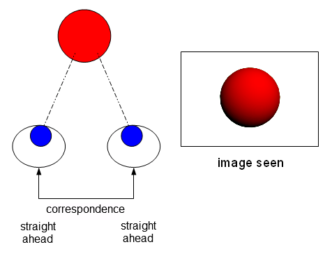
On the retina of each eye is an area called the fovea. It provides you with your clear, sharp central vision. In your brain, there is a LINK between these two foveas, and your brain operates under the assumption that the two foveas are ALWAYS pointing at the same place in space. In other words, the two foveas CORRESPOND to each other, and the brain fuses the image from each fovea into one stereoscopic image. Why does it fuse them together? Because, it is operating under the assumption that the two foveas always point to the same place in space.

This all works great when the two eyes are ACTUALLY pointing at the same thing, as in the image above. But what happens when one eye is strabismic and pointed at something else? Visual confusion or double vision results. Why? Because the brain still believes that the two eyes point at the same place in space. Thus, it fuses the images together into a crazy mess.

Going through life with double vision and visual confusion is not very easy, though. So the brain usually adapts by suppressing the image from one eye, trying to create some semblance of normal vision. The retinal link still remains intact.


There's a weirder, sneaker way that the brain can adapt, however. Break the retinal link! If the brain stops assuming that both eyes point at the same area of space, then the problem is solved. It adapts to the sad truth of reality: both eyes are NOT pointing at the same thing. Thus, each eye acts as an independent camera, with the images from both eyes being neatly pasted together in the middle.


This is ARC. The brain gives up on the notion that both eyes point at the same place in space. Instead of suppressing half of the visual field, it joins them together in the middle to create a panoramic view.

(I've read that ARC is much more common in esotropes, but I'm not sure how it works. Do they see a panorama? I'm not sure what they do...)

Anyway, back to myself. After reading that passage of Stereo Sue's book, I said "Okay, I have must have ARC. What an interesting factoid," and I didn't really think much of it. It wasn't until later, after researching it more, that I realized that it was not a good nor interesting thing. I read in Dr. Greenwald's book that Dr. Brock believed that ARC was incurable (though Dr. Greenwald himself did not believe this and had developed his own system for treating it). I also read that trying to convert ARC to NRC (normal retinal correspondence) can sometimes result in permanent double vision, and that some eye care professionals won't attempt it. And various other places, I just simply read that it was very hard to treat, with phrases like "exceedingly difficult" and "poor prognosis" being used.

At this point, I asked my optometrist and vision therapist if they thought I had ARC. Both of them didn't think that I did, and said that it was rare. My optometrist didn't think that I had it, but didn't really have the tools at the time to check if I had it or not.

So I said to myself, "I'll just figure it out myself." Here are my findings:


Josh's Findings
A report.


Hole in hand activity:
Result: Fail.

Camera flash test:
This classic ARC test involves flashing each eye with specially taped up camera flash.


-Hold it horizontally in front of one eye, stare at the piece of black tape in the middle. Flash.
-Hold it vertically in front of the other eye, stare at the piece of black tape in the middle. Flash.
-Close your eyes. Open your eyes. Look at a blank wall. Describe what you see.

When I do this test, I usually see one of the ARC versions. BUT, if I blink my eyes a lot, the two lines start moving toward each other into the NRC formation. What does that mean?!?! Fail? Pass?

Color Luster:
Wear red/green glass. Look at a light box. Try to mix the colors together. You fail if you see a half red light box, half green light box. My result: Mixed. I can see half red half green all day, but I can also make them mix a little if I squint and blink a lot.

Lid Glow:
Close one eye. Shine a penlight in your closed eye. Look at a black dot on the wall with your open eye. Try to make the black dot on the wall move into the glow you are receiving from your closed lid. Result: Pass... kind of. It's hard to do, but I can make it happen if I turn my head and blink a lot.

String test:
Wear red/green glasses. Hold a Brock string up to your nose. Fixate on a bead and make an X. If the GREEN string comes out of the RED eye, and the RED string comes out of the GREEN eye, you pass (crossed diplopia). Result: Passed with flying colors!

Bagolini Striated Lens test:
I won't go into detail about this test, but I got a weird result. I didn't get the ARC result, but I got uncrossed diplopia when I was supposed to have crossed diplopia (I think...). Result: Mystery.

Evidence for ARC:
I do a lot of things that one with ARC would do. When put on red/green glasses, relax my eyes, and look around the room, I see a half red half green room. I also feel that when doing fusion activities, I have to overcome the initial tendency to "paste" images from each eye together side by side. The first time I looked into the cheiroscope, I saw two totally separate images with no fusion. The same thing happened the first time I looked through a stereoscope, and the same thing happened when I first tried to do mirror overlap. Side by side images pasted together; panorama. I have also found it hard to gain even peripheral stereopsis, which is supposed to be "easy" (but this just changed today, see below).

Evidence against ARC:
I can also do a lot of things that one with ARC shouldn't be able to do. For example, I can make my eyes work as a team and play cards with antisuppression playing cards. I can see and play around with red/green tranaglyph slides. I can flat fuse a polarized vectogram into one image. I can do flat fusion stereoscope activities. And just today, I finally saw some depth on a polarized vectorgram! (but that's for another blog post).

Conclusion:
I believe have some ARC, but I am able to switch to NRC when doing vision therapy exercises that provide me with enough feedback (such as with anaglyphic activities and the Brock string).

I also believe that I have been self treating this ARC instinctively. When doing red/green anaglyphic activities, I often place objects that can only be seen by the RIGHT eye on the LEFT side, and objects seen by the LEFT eye on the RIGHT side. I then proceed to touch all over them with my hands to keep them "on." I have done this because it is "harder", and I now realize that it forces me to fuse the whole visual area into one image, and reinforces it with motor feedback.

If the object that can only be seen by the RIGHT eye is placed on the RIGHT, and the object that can only be seen by the LEFT eye is placed on the LEFT, then it is possible for me to "paste" the images together, even when wearing red/green glasses. Thus, the activity can be rendered useless by a smart brain if you're not careful.

I certainly hope that my tendency for pasting images together in ARC fashion isn't an insurmountable obstacle for gaining binocular stereoscopic vision. I work very hard to do my vision therapy homework and to force my eyes to work as a team. And I am usually successful. But I still have that lingering fear... What if it never happens? What if my brain is just broken?